

Multimodal Advanced Radiography Station

MARS |  CG-1D | HFIR

Mission Statement

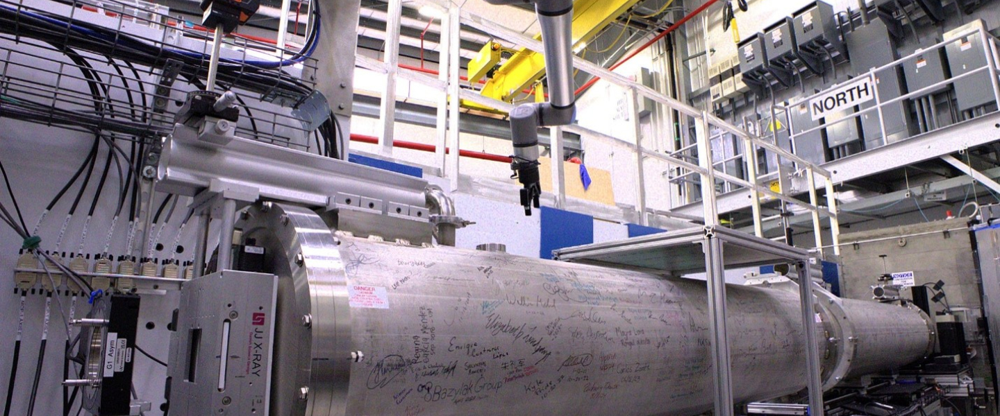
The Multimodal Advanced Radiography Station (MARS), HFIR beamline CG-1D, provides high-resolution radiography and computed tomography imaging capabilities across multiple length and time scales for diverse applications.

Instrument Description

MARS is designed as a flexible platform on which to perform high-spatial and high-temporal resolution imaging using an intense, polychromatic cold neutron beam.  In addition, white-beam neutron grating interferometry (nGI) is now offered to provide spatially-resolved neutron phase- and small-angle scattering contrasts (see Ref. 4). The instrument configuration, including beam collimation, sample platforms, sample environments, and detectors may be tailored for each measurement. The unique imaging capabilities at MARS complement those of the VENUS instrument at the Spallation Neutron Source, which is dedicated to neutron imaging with an emphasis on time-of-flight (i.e., energy-dependent) contrast. 

Instrument papers

  1. Torres et al., “Overview of MARS: the Multimodal Advanced Radiography Station at the High-Flux Isotope Reactor,” In: Craft, A.E., Bilheux, H.Z. (eds) Proceedings of the 12th World Conference on Neutron Radiography. WCNR 2024. Springer Proceedings in Physics, vol 348. Springer, Cham. DOI: 10.1007/978-3-032-15003-5_33
  2. Santodonato et al., “The CG-1D Neutron Imaging Beamline at the Oak Ridge National Laboratory High Flux Isotope Reactor,” Physics Procedia 69, pp. 104-108 (2015). DOI: 10.1016/j.phpro.2015.07.015
  3. Crow et al., “The CG1 instrument development test station at the high flux isotope reactor,” Nuclear Instruments and Methods in Physics Research Section A: Accelerators, Spectrometers, Detectors and Associate Equipment 634, no. 1, pp S71-S74 (2011). DOI: 10.1016/j.nima.2010.06.213
  4. Zhang et al., “Neutron Grating Interferometry at the High Flux Isotope Reactor,” In: Craft, A.E., Bilheux, H.Z. (eds) Proceedings of the 12th World Conference on Neutron Radiography. WCNR 2024. Springer Proceedings in Physics, vol 348. Springer, Cham. DOI: 10.1007/978-3-032-15003-5_41


Applications

MARS supports a broad range of materials research across natural and physical sciences, engineering, and advanced manufacturing. Research areas that can benefit from MARS include, but are not limited to…

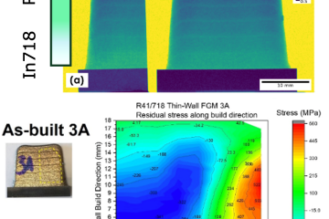
  • Additive Manufacturing - Porosity; internal structure; quantitative comparative analysis of neutron-computed tomography data with engineering drawings
  • Energy Storage and Generation - Operando ion transport in energy storage materials; three-dimensional mapping of ions in electrodes; fluid transport in fuel cells and electrolyzers
  • Nuclear Materials - In situ molten salt diffusion, solubility, and gas transport at high temperatures; inhomogeneities in nuclear fuel material
  • Transportation Technologies - Particulate deposition in vehicle parts; two-phase transport in heat pipes; multiphase constrained jet flows; metal casting; reservoir flow, creation, and production
  • Plant Systems Biology - Partitioning, transport, and fate of carbon fixed by plants; carbon biosequestration; modified bioenergy feedstock plants; cavitation and gas embolism in plants
  • Plant-Soil-Groundwater Systems - Transport and interactions of fluids in porous media; water infiltration and aquifer recharge; plant-plant and plant-fungal interactions; change in pore structure and voids after repeated thawing and freezing of permafrost soil
  • Biological, Forensic, and Medical Studies - Internal structures; mapping contrast agents; cancer research; wood and biomass pyrolysis
  • Food Science - Water migration and degradation through time
  • Archeology and Paleontology- Examination of cultural artifacts; 3D phase and structural analysis of fossils


Specifications

Wavelength Polychromatic (white beam): 0.8 < λ < 6 Å, peak at 2.6 Å, and average of 3.6 Å
Sample positions, L
(L: distance from aperture)
Downstream position: L = 6.59 m
Upstream position: L ~ 1.0 m
Collimation, L/D
(D: pinhole size)
Pinhole apertures
      Downstream: 400 – 2000
      Upstream: 60 – 300 
Slit apertures (3 mm x 20 mm) both horizontal and vertical are now available
Beam size

Downstream: 8.6 cm x 8.6 cm with slits
Upstream: ~3 cm diameter

Detectors

Charge-coupled device (CCD) and scientific complementary metal-oxide-semiconductor (sCMOS) cameras optically coupled to Zn:S or Gadox-type scintillators of various sizes and thicknesses.

More information