The enzyme PKG II (protein kinase G II – a cyclic guanosine monophosphate dependent kinase) plays an important role in human health, but can increase the risk of diseases like stomach cancer and osteoporosis if not activated. Stomach cancer kills 754,000 people globally each year and osteoporosis, which causes weakened bones, affects over 200 million people worldwide , so researchers are keen to understand how this enzyme interacts with known activators in order to guide the development of future drugs and treatments.
As part of this endeavour, researchers have proved neutron crystallography as a viable technique to gain insights into this process, using it for the first time to look at the PKG II enzyme in action.
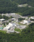
As published by a global collaboration in Biochemistry, crystals grown by researchers at the US Department of Energy’s Oak Ridge National Laboratory (ORNL) in Tennessee, US, using PKG II protein provided by the Baylor College of Medicine in Texas, US, were analysed at the Institut Laue-Langevin (ILL) in Grenoble, France.
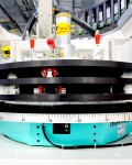
The LADI-III beamline at the ILL – capable of collecting high resolution diffraction data from very small crystals – and the IMAGINE instrument at ORNL, made it possible for researchers to critically assess the hydrogen bonding interactions during the activation of PKG II, allowing for the detailed study of the activation mechanisms that may help to prevent stomach cancerandosteoporosis. This in turn will help scientists to identify new routes for rational drug design to treat such diseases in the future.
The results of this study are also particularly compelling because neutron crystallography makes it possible to study the PKG II function at room temperature. The usual need to cool samples to cryogenic temperatures (typically 100 Kelvin, i.e. -173 Celsius) when using X-ray crystallography can alter the structure of enzymes, making it harder to confidently describe activation mechanisms and catalysis – the process controlling the speed of chemical reactions in the body.
This work has also shined light on the relationships influencing enzyme activation, with implications beyond PKG II, to other protein kinases such as protein kinase A (PKA), which is associated with cancer incidence. Comparative analysis of the backbone hydrogen/deuterium exchange patterns in PKG II and previously reported PKG I structures (see Biochemistry 2014) has suggested that the activator’s potency depends on its ability to efficiently decrease overall protein dynamics. While further studies are needed to identify how potent activators need to be to bind the strongest, if confirmed, the results will be highly significant for future drug development across a range of diseases – particularly considering ~2% of all human genes encode for protein kinases, and over half of these are linked to various diseases such as cancer and diabetes.
Matthew Blakeley, LADI-III beamline scientist at the Institut Laue-Langevin (ILL) and co-author of the study, said: “Neutron crystallography allows us to determine the positions of the hydrogen atoms providing us with crucial information on the hydrogen-bonding interactions between small molecule drugs and their protein targets. Moreover, the ability to distinguish between hydrogen and deuterium with neutrons provides us with information relating to the binding dynamics of the protein-drug complexes, which may be critical for the potency of activators of PKG I and PKG II.”
Andrey Kovalevsky, IMAGINE instrument scientist at Oak Ridge National Laboratory and senior co-author of the study said: “It is exciting that we have been able to observe the information transfer across the PKG II protein during the activation process and begin to establish what might be influencing some small molecules to activate the enzyme better than others. This knowledge will have a great impact on our understanding of various diseases today, and will help us to develop the drugs and treatments of the future.”
Professor Choel Kim, associate professor at Baylor College of Medicine and co-author of the study said: “While understanding PKG signaling and cellular functions requires inhibitors and activators with high selectivity and potency, such chemical tools are not currently available. Improving selectivity and potency of known compounds and identifying new effector molecules would be an obvious strategy to resolve this challenge, but this requires molecular-level understanding of how these compounds interact with different functional domains of PKG. The neutron structure presented here shows PKG II-activator interaction in unprecedented atomic detail, and thus serves as a solid starting point for the rational design of PKG II selective activators.”- Original Post from ILL
About ILL – the Institut Laue-Langevin (ILL) is an international research centre based in Grenoble, France. It has led the world in neutron-scattering science and technology for almost 40 years, since experiments began in 1972. ILL operates one of the most intense neutron sources in the world, feeding beams of neutrons to a suite of 40 high-performance instruments that are constantly upgraded. Each year 1,200 researchers from over 40 countries visit ILL to conduct research into condensed matter physics, (green) chemistry, biology, nuclear physics, and materials science. The UK, along with France and Germany is an associate and major funder of the ILL.
About ORNL – Oak Ridge National Laboratory (ORNL) operates two neutron source facilities, the High Flux Isotope Reactor and the Spallation Neutron Source. Built and funded by the U.S. Department of Energy (DOE) Office of Basic Energy Sciences (BES), the two facilities combined house 30 neutron scattering instruments, providing researchers with unmatched capabilities for understanding the structure and properties of materials, macromolecular and biological systems, and the fundamental physics of the neutron. More than 1,200 unique users from around the world use ORNL’s neutron sources annually. ORNL is managed and operated by UT-Battelle for DOE. For more information visit http://science.energy.gov


