

Contacts
-

Instrument Scientist
-

Instrument Scientist
-

Instrument Scientist
")
Biochemistry at the atomic scale
Understanding essential biochemical processes at the atomic level is the key to discoveries in the bioenergy, biomedical, and pharmaceutical sciences. IMAGINE’s high resolution allows researchers to study proteins and other biomacromolecules, including those involved in processes such as biofuels production, the development of disease, and drug design..
CG-4D’s IMAGINE instrument is a state-of-the-art, neutron image plate, single-crystal diffractometer that provides atomic resolution information on inorganic, organic, metallo-organic, and macromolecular single crystals that enables their chemical, physical, and biological structure and function to be understood. IMAGINE benefits communities with interest in pharmaceuticals, minerals and materials, small molecules, molecular organo-metallic complexes and metal-organic frameworks and enables the neutron crystal structure of oligonucleotides and proteins to be determined at near atomic resolutions (1.5 Å).
Macromolecular structure and function
Supra-Molecular Crystallography
Sample Environment Under Development: Materials under Extreme Environment
| Flux | ~107 n/s/cm2 |
| Cross section | 2.0 x 3.2 mm |
| Wavelengths minimum | 2.0, 2.8, 3.3 Å |
| Wavelengths maximum | 3.0, 4.0, 4.5 Å |
| Detector | Neutron image plate (Gd2O3 doped BaF(Br.I):Eu2+) |
| Detector size | 1200 x 450 mm |
| Pixel size | 125, 250, 500 µm |
| Sample-to-detector distance | 200 mm |
| Goniometer | Kappa and phi rotation axes |
| The calculator assumes that only one crystal grows in the drop. Protein solubility is not considered. Accuracy of protein concentration is essential. |
| O’Dell W.B., Bodenheimer A., Meilleur F. (2016) Neutron protein crystallography: A complementary tool for locating hydrogens in proteins. Arch Biochem. Biophys. 602:48-60 |
Instrument Scientist
Instrument Scientist
Instrument Scientist
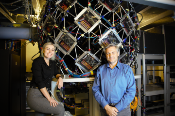
 and Dean Myles at the High Flux Isotope Reactor’s IMAGINE instrument for protein crystallography, which will be significantly upgraded through DOE’s BRaVE initiative for biopredaredness. Credit: Jeremy Rumsey/ORNL, U.S. Dept. of Energy")


Oak Ridge National Laboratory is managed by UT-Battelle LLC for the US Department of Energy