Neutron imaging (i.e., neutron radiography with computed tomography) is a noninvasive, nondestructive technique that measures static or time-dependent features of objects under real-world operating conditions, like pressure, temperature, and tensile and compression loading. Attenuation-based neutron imaging works by measuring a shadow radiograph of an object, much like x-ray radiographs. Advanced techniques use the phase, small-angle scatter, or spin of neutrons as a contrast mechanism. Neutrons are a complementary probe to protons and x-rays in their interactions with matter because they penetrate most metallic materials while being highly sensitive to lighter elements such as hydrogen or lithium.
A useful research technique across most scientific disciplines—from materials science, physics, geosciences, and engineering to plant physiology, biology, archeology, and forensics—neutron imaging provides information about the structure of materials and objects that cannot be easily achieved with other techniques. Neutron imaging at the High Flux Isotope Reactor Cold Neutron Imaging Facility has been used for in situ real-time measurement of lithium transport in commercial batteries, water transport in plant roots, soot deposition in particulate filters, rapid imbibition of water in fractured sedimentary rocks, two-phase flows in refrigerants, and many other studies. Imaging at the Spallation Neutron Source has included mapping crystalline information for additively manufactured metals and isotopic content of heavy elements for nuclear materials.
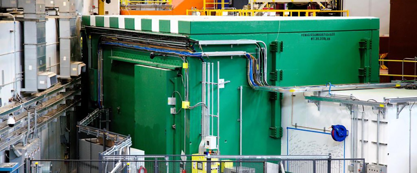
Two neutron imaging instruments are available at ORNL and there are ongoing efforts to build a third instrument, the VENUS imaging beamline at the Spallation Neutron Source. VENUS will provide a dedicated imaging instrument for neutron wavelength-dependent measurements.
For more imaging information please visit: https://neutronimaging.pages.ornl.gov/en/


