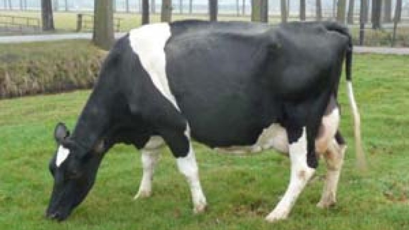
Martha, a cow placidly grazing in a field in The Netherlands, became an important collaborator with researchers who successfully analyzed and characterized the internal protein structure and the composite particles of her milk using small-angle neutron scattering at ORNL’s High Flux Isotope Reactor (HFIR).
Casein micelles, a family of related phosphorus-containing proteins, make up 80% of the protein in cow milk. They are the building blocks of dairy products such as yogurt and cheese, supplying amino acids, calcium, and phosphorus to the body. More important, they are the principal vehicle for delivering calcium phosphate to rapidly growing newborns.
Researchers have long struggled with the challenge of unraveling the internal structure of this protein. An international collaboration was formed among University of Utrecht researchers C. G. (Kees) de Kruif and Andrei Petukhov; Volker Urban, an instrument scientist at HFIR; and Thom Huppertz from NIZO, a Dutch independent contract research company that helps food and ingredient companies improve the quality and profitability of their products.
The researchers used the General-Purpose Small-Angle Neutron Scattering instrument (GP-SANS) to study samples of milk from Martha, a cow on the de Kruif family’s farm. They compared the neutron scattering data with various theoretical models of casein structure that have been proposed in the literature. The results showed that one model prevails: the casein micelle proteins are composed of a protein matrix in which calcium phosphate nanoclusters (about 300 per casein micelle) are dispersed.
Neutron scattering also revealed that the protein’s matrix has fluctuations in density that are attributable to the hydrophobic (water-repelling) interactions of the casein proteins.
The researchers collected critical parameters for their experiments by first measuring the properties of casein micelles of individual cows to obtain data for size, size distribution, protein composition, density, and water content, which served as input.
“Small-angle neutron contrast variation is the tool of choice to solve the problem,” explained de Kruif, “because one can calculate the scattering intensity with no ambiguity.”
“We needed a SANS instrument for two reasons. The length scale involved is from a few tenths of a nanometer to a few hundred nanometers. Only neutrons can do such an in situ contrast variation for testing the various models.”
“Furthermore, the casein micelle models show a distribution of mass within each particle. Given the parameters determined independently, one can calculate the neutron scattering intensity. Only if a model proposes the correct distribution of matter can one calculate all the scattering intensities self-consistently.”
“We used the casein micelles of a single cow (Martha) because the micelles were somewhat smaller than average and less poly-disperse. This further limits the freedom of the parameters. We showed that these samples were representative for the whole milk.”
The work may have application one day in other areas, de Kruif said. “Casein micelles can be used as delivery vehicles for various compounds, such as vitamins and minerals. Knowing the internal structure will be useful in developing such applications.”
The team has since applied for additional beam time to study the initial stages of yogurt and cheese making, both a specialty of The Netherlands.
This research was funded by the U.S. Department of Energy (DOE) Office of Basic Energy Sciences, DOE Office of Biological and Environmental Research, Utrecht University, The Netherlands, and NIZO food research.
Published Work
de Kruif, C. G., Huppertz, T., Urban, Volker S., and Petukhov, A. V., “Casein micelles and their internal structure,” Adv Colloid Interfac, 171-172, 36-52 (2012), doi: 10.1016/j.cis.2012.01.002.


