Andrey Kovalevsky

Andrey Kovalevsky

Contact: kovalevskyay@ornl.gov or (505) 310-4184

Research

Living organisms depend on the action of enzymes that catalyze chemical reactions too slow to support life. Enzymes are central in fighting disease, with most current clinical drugs targeting human, bacterial, or viral enzymes. In biotechnology, utilization of enzymes is the “green” option for biofuel and chemical production. Understanding the remarkable catalytic efficiency of enzymes in mechanistic terms has been the goal of biochemistry for decades, and the knowledge has had profound effects on biotechnological processes and drug design. Science is still working toward this elusive goal, demanding understanding at the level of atoms and electrons. Our approach to study enzymes combines innovative experimental and theoretical methodologies, including X-ray and neutron crystallography, neutron spectroscopy, and molecular simulations, in a novel way, providing mechanistic views of enzymes with unprecedented detail and then applying this knowledge to protein engineering and drug design.

Enzymes rely on moving hydrogen (H) atoms and activating water molecules for their function. X-ray crystallography and nuclear magnetic resonance (NMR) — the major techniques for studying protein structure and function — are typically incapable of producing necessary information on the location and movement of H. A critical limitation of X-ray crystallography is the weak sensitivity of X-rays to H atoms. With one electron, H is hard to observe in X-ray structures. H often remains invisible even at ultra-high resolution, so the protonation states of amino-acid residues and ligands remain unknown. Incorrect inference of H positions in protein structures often leads to significant gaps in our knowledge of how they function. Neutrons are scattered as strongly by H, and its heavier isotope deuterium (D), as they are by other elements in proteins. Consequently, neutron crystallography provides a direct means for accurate determination of the locations of H in a macromolecular structure even at medium resolutions of ~2.5 Å. Yet, H possesses negative neutron scattering length, causing cancellation effects in the nuclear density maps, and also has a strong incoherent scattering component that contributes heavily to background. Thus, H is usually exchanged with D in protein crystals to increase signal-to-noise ratio and improve neutron diffraction. When both techniques are combined in the X-ray/neutron (XN) methodology and macromolecular structures are refined simultaneously against both diffraction datasets, better, more complete and accurate protein structures are obtained. These crystal structures, however, represent only the snapshots of an enzymatic reaction, providing details on intermediate states and possible H transfer routes along the reaction pathway. But, crystallography cannot offer the structures of fleeting transition states, nor can it map reaction pathways. Quantum chemistry in combination with molecular mechanics and molecular dynamics can map a reaction pathway in a protein; however, an accurate theoretical result can be achieved only if we precisely know the H atom positions.
 
Our research program consists of three major projects:
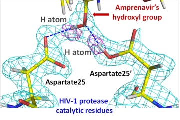
  1. Mechanistic, drug binding and drug resistance studies of the human immunodeficiency virus type 1 (HIV-1) protease and its precursors.
  2. Mechanistic studies of reactivation of organophosphate-conjugated human acetylcholinesterase (hAChE) by oximes and the design of novel accelerated reactivators.
  3. Mechanistic studies of vitamin B6 (PLP)-dependent enzymes.
 
Project 1. HIV-1 protease enzyme plays a vital role in the viral replication cycle during maturation of a newly emerged viral particle into an infectious virion. The protease is expressed as part of the Gag-Pol polyprotein, and catalyzes hydrolysis of the peptide bonds within Gag and Gag-Pol polyproteins for its own maturation (called autoprocessing) and to generate other mature viral enzymes and structural proteins. HIV-1 protease has proven to be an effective target for drug design and development, with 9 clinical protease inhibitors currently marketed in the U.S. It has also been viewed as one of the best examples of the structure-guided drug design. However, the long-term potency of protease inhibitors is thwarted by rapid emergence of drug resistant protease variants, which necessitates the constant development of new drugs active against resistant protease variants, with higher barrier to resistance. Understanding HIV-1 protease function and the mechanisms of drug resistance at atomic level is of paramount importance in our ability to design improved drugs. We are using X-ray and neutron crystallography, neutron spectroscopy and molecular simulations in order gain true atomic picture of the enzyme function, drug binding, and to identify and quantify drug resistance mechanisms. Our neutron structures of HIV-1 protease with clinical drugs illustrated, for the first time, the exact non-covalent interactions that lead to the effectiveness of amprenavir and darunavir clinical HIV-1 protease inhibitors on the wild-type and mutant enzyme. They also underscored the shortcomings of obtaining crystallographic structures with X-rays at low temperature. Neutron spectroscopy experiments have shown how the enzyme’s global dynamics, or vibrations, affect drug binding, that may lead to the identification and quantification of a new drug resistance mechanism on the molecular level. These studies are producing one-of-a-kind, valuable information for the design of new protease inhibitors with improved bindig to drug resistant HIV-1 protease drug resistant variants. In the future, we are aiming to look at extremely drug resistant protease variants with both neutron diffraction and spectroscopy. We also are gearing up at studying the enzyme’s autoprocessing – the one step in the HIV-1 replication cycle not targeted for drug development.
 
Project 2. The world community is presently witnessing an unfortunate insidious use of nerve agent organophosphates (OPs) as chemical weapons. Highly efficient antidotes to protect population against those anti-acetylcholinesterase (AChE) poisons are not available. The concept of nucleophilic reactivation of OP-inhibited AChE emerged in 1950s from the monumental studies of Irwin Wilson and colleagues who showed that hydroxamates, oximes and hydroxylamines could reactivate alkylphosphate-inhibited AChE. Subsequent studies over the past five decades have yielded minor improvements in reactivation rates and a moderately enhanced efficacy for the antidotes, such as HI6 and MMB4. Given the rapid AChE inhibition by nerve agent and pesticide OPs, and far slower nucleophilic reactivation by oximes, challenges and opportunities remain. This project aims to investigate structural limitations for the oxime reactivation by cutting edge biophysical study of the native and OP inhibited AChE in order to design accelerated reactivators devoid of those limitations. The objective of this collaborative translational endeavor with investigators at the University of Utah, University of California, San Diego, and Oak Ridge National Laboratory funded by the NIH’s COUNTERACT program is directed to developing a new generation of accelerated oxime reactivators of nerve agent OP-inhibited human acetylcholinesterase (hAChE). We are performing cutting edge biophysical studies to understand how slow molecular motions, protonation and ionization states in the mechanism of oxime reactivation of nerve agent OP-inhibited hAChE influence and in effect limit its efficacy. Only neutron diffraction can resolve protons critical for analysis of both reactivation and catalysis. Fort this purpose we are working with NASA’s CASIS program in order to grow neutron-quality crystals on the International Space Station in microgravity. Current AChE structures determined by X-ray crystallography are becoming limiting in the design of efficient antidotes. These data will be critical for timely development of accelerated oxime reactivator antidotes. Our low- and first-ever room-temperature X-ray structures of apo- and VX-conjugated hAChE in complex with experimental oximes have demonstrated the mode of binding of these reactivators to the enzyme’s active site and provided insights into how they reactivate the enzyme. Also, using small-angle X-ray scattering we have shown that hAChE inhibition by pesticide POX leads to the disruption of the enzyme’s dimeric structure, whereas neutron spectroscopy experiments demonstrated that POX results in softening of the vibrational flexibility of the enzyme that perhaps results in the disruption of the dimer. In addition, molecular dynamics simulations are probing the modes of oxime binding supplementing structural results.
 
Project 3. Pyridoxal phosphate (PLP), the enzyme cofactor derived from vitamin B6, catalyzes a myriad of reactions involving a-aminoacids. Dependent on the various active site environments, the products released are the result of deaminations, decarboxylations, b and g eliminations, racemizations, and retro-aldol cleavage. Our interest is to understand how hydrogen-bonding networks, unique to each type of reaction, modulate PLP activity. We focus on comparing aminotransferase activity to the b- and g- elimination process. The activities of all PLP dependent enzymes involve the formation of a Schiff base between the PLP C4’ aldehyde and the e-amine of an active site lysine (internal aldimine), which subsequently transfers to substrate (external aldimine). In most proposed mechanisms, protonation of the pyridine nitrogen and/or the Schiff base nitrogen promotes bond rearrangement invoking substrate transformations. Also, the PLP phenolic oxygen (O3’) presumably forms an internal hydrogen bond with the Schiff base nitrogen and in some cases donates a hydrogen to form an oxyanion that directs bond rearrangement. Modulation of the various PLP activities and stabilization of intermediates is derived from hydrogen bonding of PLP and substrates to enzyme active site moieties and structural waters. We aim to determine if and when these proposed PLP protonation events occur and to discern these bonding networks establishing a better understanding of PLP mechanisms. Interestingly, all proposed mechanisms invoke the deprotonation of single-bond amino groups so that they become nucleophiles. With single-bond nitrogens of substrates having basic pKa’s and double-bond nitrogens of the PLP and Schiff base having acidic pKas, these protonation and deprotonation steps require assistance. We are studying several PLP-dependent enzymes – aspartate aminotransferase, tryptophan synthase and threonine synthase.