
Probing the Invisible: Neutrons Illuminate Ligand Structures in Quantum Dots
July 15, 2025
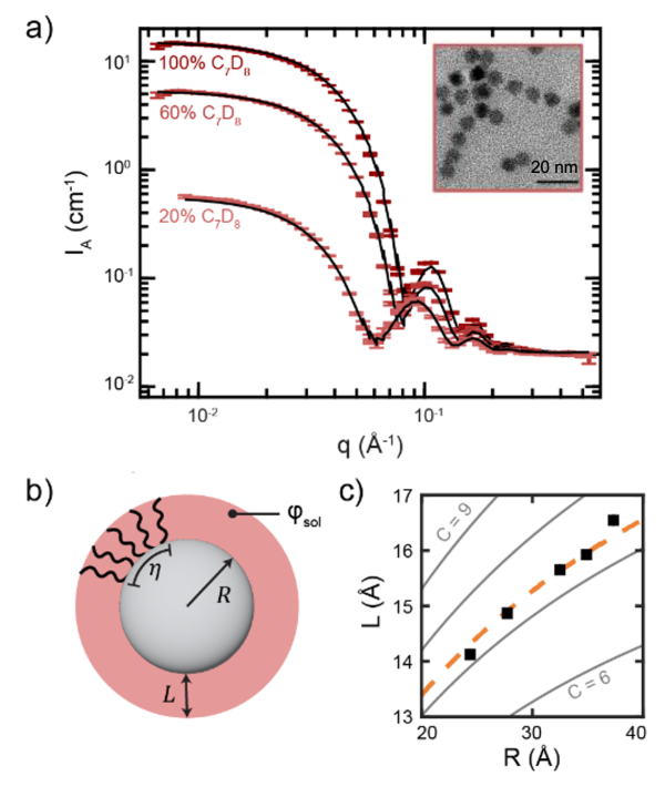
SANS data illustrates a positive correlation in quantum dot core size (R) and ligand shell thickness (L). a) SANS data at 100%, 60%, and 20% toluene solvent deuteration (colored data points with error bars) and core-shell model fits (black lines) for 7.4 nm diameter PbS quantum dots. Inset: TEM of PbS quantum dots.
Scientific Achievement
Precise ligand shell thickness of colloidal quantum dots was revealed by small-angle neutron scattering (SANS), providing essential information unattainable by other techniques.
Significance and Impact
Deepened understanding of ligand shell thickness enables improved control of the self-assembly and functionality of quantum dots, critical for applications in optoelectronics, sensing and photocatalysis.
Research Details
- Organic ligand shells of quantum dots were characterized by SANS; complementary x-ray scattering revealed interparticle interactions and self-assembled structure.
- Ligand shell thickness was influenced by both the quantum dot core size and the type of solvent.
“Ligand Shell Thickness of Colloidal Nanocrystals: A Comparison of Small-Angle Neutron and X‑ray Scattering,” J. Am. Chem. Soc. 147, 13859-13870 (2025)
doi:10.1021/jacs.5c02070