

Protein Structures Involved in Cancer Cell Proliferation Revealed
May 15, 2025
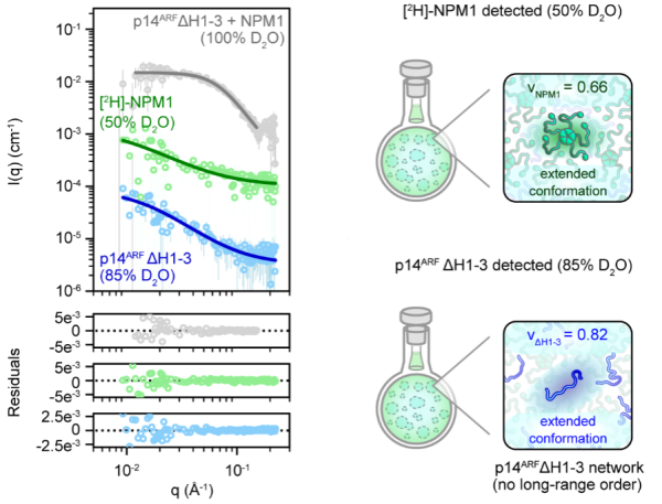
Small-angle neutron scattering data (top left) and modeling residuals (bottom left) for isotopically-labeled NPM1 and p14ARF measured under the contrast conditions are shown on the right. The protein structures were revealed in the highly concentrated gel-like state that exists when the two proteins interact and phase separate.
Scientific Achievement
The interactions between two proteins, NPM1 and p14ARF, involved in cellular proliferation were revealed.
Significance and Impact
The two intrinsically disordered proteins phase separate into disordered gels when they bind. The protein structures that were not accessible by other experimental techniques provide new insight into cancer cell proliferation regulation.
Research Details
- Deuterium labeling
- Small-angle neutron scattering with contrast variation
- Nuclear Magnetic Resonance spectroscopy
“p14ARF forms meso-scale assemblies upon phase separation with NPM1”. E. Gibbs, Q. Miao, M. Ferrolino, R. Bajpai, A. Hassan, A. H. Phillips, A. Pitre, R. Kümmerle, S. Miller, G. Nagy, W. Leite, W. Heller, C. Stanley, B. Perrone and R. Kriwacki, Nat. Commun. 15, 9531 (2024). https://doi.org/10.1038/s41467-024-53904-z