

Revealing Enzymes Cluster in Metal–Organic Framework (MOF) Nanocages
June 15, 2026
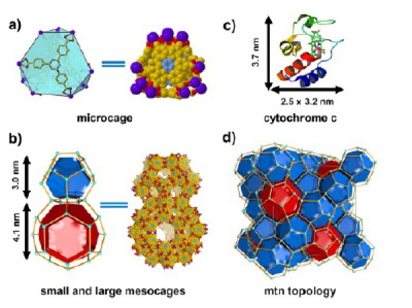
Structure of Tb-mesoMOF a) microporous truncated tetrahedron; b) two connected mesopores (blue represents small mesopore and red represents large mesopore); c) The structural dimensions of Cyt. C; and d) mtn topology of Tb-mesoMOF.
Scientific Achievement
Direct observation of cytochrome c clustering inside crystalline Tb-mesoMOF, exposing enzyme arrangements that are difficult to access with conventional techniques.
Significance and Impact
The work shows neutrons can non-destructively probe the internal structure of porous crystals, helping design MOF biocatalysts with higher enzyme loading, stability, and function.
Research Details
- Contrast matching (60% D2O/H2O ) minimized the MOF signal and isolated Cyt. c scattering.
- A low-q feature indicated enzyme clustering; 50 mM Tris produced a ~59 nm superstructure.
- UV-vis, FTIR, PXRD, and N2 sorption linked buffer chemistry to loading kinetics and pore evolution.
“Monitoring Enzyme Clustering in a Highly Crystalline Metal–Organic Framework by Small-Angle Neutron Scattering”, Angewandte Chemie. Internatianal Edition 65, e22967 (2026).
https://onlinelibrary.wiley.com/doi/10.1002/anie.202522967